International Journal of Scientific and Research Publications
Monograph:
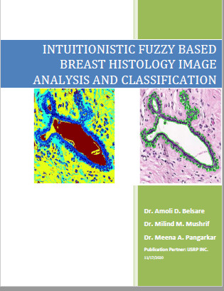
Title: INTUITIONISTIC FUZZY BASED BREAST HISTOLOGY IMAGE ANALYSIS AND CLASSIFICATION
Author/s: Dr. Amoli D. Belsare, Dr. Milind M. Mushrif, Dr. Meena A. Pangarkar
PREFACE
In this digital era, everyone is acquainted with the use of digital devices and many people use them every day. Use of digitized data has risen exponentially in the healthcare sector too & is used extensively in diagnostics, treatment & post treatment follow-ups. But there is still a vast scope for further advancements and refinements. Keeping this in mind, the present work focuses on digitizing pathological examination of a biopsy sample from a patient suspected to have Breast cancer. The study aims to be helpful in reducing the workload of the pathologist as well as help in decision making by reducing inter- and intra- observer variability.
Breast cancer is second largest cancer in India which results in a lot of mortality and morbidity in the young and productive age group women. It is a treatable and curable cancer if diagnosed and treated early, resulting in saving a lot of lives.
Thus, digitization of the microscopic images of biopsy samples was done so as to provide a helping hand to pathologist to save time and subjectivity of diagnosis. The multiple microscopic images were analyzed by use of image processing techniques and results were generated as positive or negative for Cancer. This tool can be used as a second opinion by pathologists/oncologists.
Briefly, a spatio-texture based image segmentation algorithm is developed for identifying breast duct accurately. This work is further extended by using intuitionistic fuzzy based approach to identify breast duct from digitized biopsy sample with more segmentation accuracy. Due to different staining concentrations at different labs, tissue image may have color variation and hence normalization is needed while preprocessing the image. Intuitionistic fuzzy considers the uncertainty of a pixel information and the normalization step in processing histology image can be avoided. Once the duct from breast tissue images is identified, basic binary classification of images as malignant and non-malignant has been performed & the results are compared with the state of art approaches from image processing. This work is very special as it involves the guidance from medical professionals in development of algorithms as well as development of image database for the pilot study along with ground truth data for evaluation.
I would like to thank all who directly or indirectly helped me in the development of the methods with guidance and support. I also extend my heartfelt thanks to my guide Dr. M. M. Mushrif & co guide from GMCH Nagpur Dr. Mrs. M. A. Pangarkar who constantly supported me in whole process of design, development of concept to publishing this book. At the end, I would like to thank my family members, Mother, Father, Father-in-law, Late Mother in law, my husband and my son who encouraged me constantly to work hard and supported me in everything without which this writing can't be completed.

 Title: INTUITIONISTIC FUZZY BASED BREAST HISTOLOGY IMAGE ANALYSIS AND CLASSIFICATION
 [READ Thesis]